Thyroid Ultrasound for Papillary Thyroid Cancer

Thyroid ultrasound is the best and most common method for evaluating the thyroid. This imaging test uses sound waves to get pictures of the thyroid gland, surrounding tissue and structures, and the lymph nodes in the neck. The first imaging study used in work-up and evaluation of papillary thyroid cancer and thyroid problems should always be an expert, skilled, high-resolution thyroid and neck ultrasound.
Thyroid Ultrasound for Papillary Thyroid Cancer
During your thyroid ultrasound for papillary thyroid cancer, a small, wand-like instrument called a transducer is placed on the skin in front of your thyroid gland. This transducer gives off sound waves and picks up the echoes as they bounce off the thyroid (and other underlying neck structures). The echoes are converted by a computer into a black and white image on a computer screen. The resolution, or ability of the machine to obtain crisp, clear pictures of even microscopic nodules, cancer, or lumps in the thyroid, is very important.
Thyroid Ultrasound for Papillary Thyroid Cancer: NO radiation
An expert ultrasound for papillary thyroid cancer exposes the patient to absolutely no radiation. Unlike a CT scan or x-ray study, ultrasound does not use radiation to obtain pictures of the inside of your body. Ultrasound uses harmless sound waves. When a patient needs imaging to evaluate their papillary thyroid cancer or other thyroid conditions, an ultrasound of the neck should always be the first imaging test. Ultrasound uses sound waves to create pictures inside your neck that allow for evaluation of the thyroid gland, structures nearby (windpipe, esophagus, blood vessels, etc.), and lymph nodes.
Again, you are not exposed to any radiation during this test. Below is a picture of a high-resolution ultrasound machine.

To find out more about how ultrasound is used to diagnose thyroid, and even parathyroid disease, please visit www.thyroidcancer.com and www.parathyroid.com.
Thyroid Ultrasound for Papillary Thyroid Cancer: Best imaging method for evaluating thyroid
As noted above, thyroid ultrasound is the best imaging method to evaluate papillary thyroid cancer. When there is concern for papillary thyroid cancer (or other thyroid disease) a skilled, thorough ultrasound needs to be done first. If your doctor orders a CT scan, MRI, or PET/CT scan to initially evaluate for papillary thyroid cancer, you need a second opinion from an expert.
Thyroid ultrasound can determine if papillary thyroid cancer is likely. This test can also be used to check the number and size of thyroid nodules and can even reveal what the blood supply looks like to these nodules. Additionally, this method is excellent to look for thyroid cancer that has spread to lymph nodes in the neck. Furthermore, ultrasound is used to perform needle biopsies of thyroid lumps, nodules, and lymph nodes to diagnose papillary thyroid cancer.
.png)
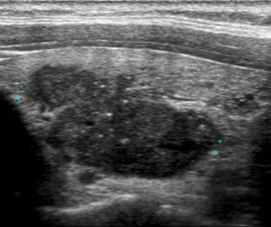
Figure 1: Ultrasound image of a cancerous thyroid nodule
.png)
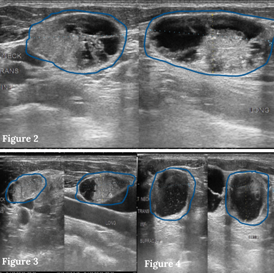
Figures 2-4: Ultrasound images of multiple enlarged, cancerous lymph nodes with a speckled, irregular appearance in the left side of the neck.
For more information about ultrasound and the diagnosis of thyroid cancer, please visit https://www.5-best-ways-to-diagnose-thyroid-cancer.
Thyroid Ultrasound for Papillary Thyroid Cancer: Need to examine lymph nodes in the neck
You should feel good knowing a thyroid ultrasound was the first imaging test ordered by your doctor to examine and diagnose papillary thyroid cancer. If you do not get a skilled, expert ultrasound done with a high-resolution ultrasound machine, however, you run the risk that some thyroid problems, particularly thyroid cancer, might be missed.
Every thyroid ultrasound must include examination of the lymph nodes and the tissues that surround the thyroid. This should be done in the middle of the neck, and on both sides of the neck, from the jaw down to the sternum (chest bone) and collarbone. Otherwise, thyroid cancer that has spread will be missed. Thus, the goal of the thyroid ultrasound is NOT just to look at the thyroid. A high-quality thyroid ultrasound also examines all the tissues on both sides of the neck to look for enlarged or abnormal- appearing lymph nodes.
High- resolution ultrasound has a detection capability as small as 1-2 mm (the size of a tip of a ball point pen) for a papillary thyroid cancer, including cancer that has spread to lymph nodes in the neck. An excellent, thorough ultrasound will determine if the papillary thyroid cancer is present or if a needle biopsy needs to be performed to diagnose thyroid cancer that has spread to the lymph nodes. The only weakness of ultrasound is that it cannot distinguish cancerous lymph nodes from inflammatory or reactive lymph nodes. Both can have very similar appearances, but ultrasound- guided needle biopsy will provide the necessary microscopic information to confirm or rule out a diagnosis of papillary thyroid cancer.
To have an expert ultrasound and thyroid evaluation done, please visit www.thyroidcancer.com/become-a-patient.
Thyroid Ultrasound for Papillary Thyroid Cancer: Expert ultrasound team is crucial
Ultimately, the quality of the ultrasound is dependent upon four factors. Each factor is critically important and listed below.
- The quality of the ultrasound machine
- The device that is held in the hand of the technologists producing the sound waves (the transducer)
- The experience and the skill of the ultrasound technologists
- The experience of the surgeon or radiologist who is interpreting the study.
The best thyroid ultrasounds for papillary thyroid cancer are done by experts who perform many on a daily basis. Your ultrasound should be performed by someone who is specifically dedicated to the ultrasound examination of the thyroid and neck.
Experience means everything when you are considering the sensitivity and accuracy of a neck ultrasound for papillary thyroid cancer. At our thyroid hospital, the ultrasound technologists only perform ultrasounds of the thyroid and neck every day.
.png)
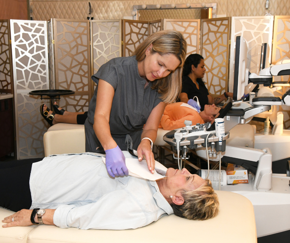
Figure 5: Our expert team of thyroid ultrasound technologists holds free thyroid screening events throughout the local community. Learn more about the thyroid ultrasound screening program.
You can’t diagnose what you cannot see or find. You also need to know what you are looking at to properly diagnose papillary thyroid cancer. This expertise only comes with tens of thousands of ultrasounds and thorough evaluation/reading of the ultrasound pictures. Settling for anything less than a highly-experienced and adept ultrasound team can be detrimental to your health when dealing with papillary thyroid cancer.
To learn more, please visit www.thyroidcancer.com and check out our blogs about thyroid cancer, thyroid problems, and treatment at www.thyroidcancer.com/blogs
Thyroid Ultrasound for Papillary Thyroid Cancer: Summary
Thyroid ultrasound is a harmless, non-invasive imaging modality that does not expose the patient to any radiation. Thyroid ultrasound is the best imaging test to diagnose and evaluate papillary thyroid cancer. When there is concern or suspicion for papillary thyroid cancer, a skilled, high-resolution ultrasound of the thyroid, surrounding structures, and lymph nodes in the neck should be done first (NOT a CT scan or MRI). Ultrasound is also used to perform guided needle biopsies of thyroid nodules and lymph nodes to diagnosed papillary thyroid cancer. Finally, a skilled, experienced, and expert ultrasound team along with a high-resolution ultrasound machine to evaluate papillary thyroid cancer is a must.
Additional Resources
- Become our patient at www.thyroidcancer.com/become-a-patient
- Learn more about the The Clayman Thyroid Center at www.thyroidcancer.com
- Learn more about our specialized Hospital for Endocrine Surgery
- Learn more about our thyroid ultrasound screening program for the Tampa community.